Cutting-Edge Advances Transforming Eye Cancer Surgery in 2025
Explore the newest surgical techniques, imaging tools, and AI‑driven approaches that are reshaping treatment for ocular tumors, from melanoma to retinoblastoma.
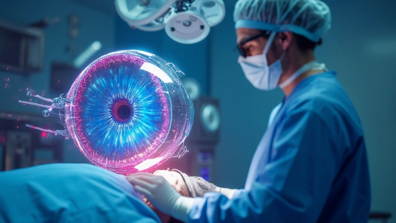
When talking about intraocular brachytherapy, a form of internal radiation that targets eye tumors by placing a radioactive source directly on the eye surface. Also known as plaque brachytherapy, this technique lets doctors focus high‑dose radiation on the cancer while sparing healthy tissue. It’s a key tool in ocular tumor, abnormal growths inside the eye that can threaten vision and even life and relies on radioactive isotopes, materials like Iodine‑125 or Palladium‑103 that emit low‑energy gamma rays suitable for eye treatment. This combination of precise placement and controlled radiation makes the procedure uniquely effective for preserving sight.
The process starts with plaque therapy, a small, gold‑backed disc that holds the radioactive isotope and is sutured onto the sclera (the white part of the eye). The plaque stays in place for a few days to weeks, delivering a calculated dose directly to the tumor. This method requires meticulous planning because the radiation must be strong enough to kill cancer cells but gentle enough to keep the retina, optic nerve, and other structures safe. Once the prescribed dose is reached, the plaque is removed and the eye begins to heal.
Because the radiation source is right next to the tumor, intraocular brachytherapy encompasses three critical goals: (1) targeted dose delivery – the isotope’s energy is focused on the lesion; (2) minimized exposure – surrounding tissues receive only a fraction of the dose; and (3) preservation of vision – many patients retain useful sight after treatment. Studies show that using Iodine‑125 in a 5‑mm plaque can control most choroidal melanomas while keeping visual acuity at acceptable levels.
Choosing the right candidates is another piece of the puzzle. Eye oncologists assess tumor size, location, and thickness, often using ultrasound and MRI, to decide if brachytherapy offers a better outcome than enucleation (removal of the eye) or external beam radiation. Younger patients, those with peripheral tumors, and individuals who value eye preservation are typical candidates. Side effects can include temporary swelling, cataract formation, or radiation retinopathy, but modern planning software helps keep these risks low.
In practice, intraocular brachytherapy influences the broader field of radiation oncology, the medical specialty that uses ionizing radiation to treat cancer. Techniques developed for the eye, such as precise dosimetry and custom plaque design, often translate to other cancers where localized dose delivery is crucial. Patients who undergo this treatment also benefit from a multidisciplinary team that includes ophthalmologists, radiation physicists, and visual rehabilitation specialists.
Below you’ll find a curated list of articles that dive deeper into each facet of intraocular brachytherapy – from isotope selection and plaque construction to patient experiences and long‑term outcomes. Whether you’re a patient, a caregiver, or a healthcare professional, these resources will give you practical insights and up‑to‑date guidance on this specialized eye‑preserving therapy.
 25 Sep 2025
Explore the newest surgical techniques, imaging tools, and AI‑driven approaches that are reshaping treatment for ocular tumors, from melanoma to retinoblastoma.